Cross Section Of A Bone Labeled : Tooth Cross Section Diagram Archer Dental / Characterization of eight different tetracyclines:
Cross Section Of A Bone Labeled : Tooth Cross Section Diagram Archer Dental / Characterization of eight different tetracyclines:. • learn about the materials that make up bone • label a cross section of bone. Related posts of cross section of a long bone bones in neck diagram. Bone marrow is the soft, highly vascular and flexible connective tissue within bone cavities which serve as the primary site of new blood cell production or hematopoiesis. The present study initially focuses on the cortex of a single sample of human bone. A cross section of a human long bone.
This colored scanning electron micrograph (sem) is showing the internal structure of a broken finger bone. Hope you enjoy and please. Tibiae (triangular cross section) showed lower stiffness values, as evident by the more levelled curves. Red marrow fills the spaces in the spongy bone. As the names suggest compact bone looks compact and the spongy bone looks like skull bone is a flat bone.
Bodypartchart Cross Section Of The Foot Labeled Anatomical Charts from cdn3.volusion.com Draw and label a cross section of a bone. Two types of bone tissues in cross section of a long bone : The wider section at each end of the bone is called the epiphysis (plural = epiphyses), which is filled with spongy bone. Virtual bone labwe need our bones to walk, run, jump and move, but this is not all they do. Hope you enjoy and please. At the outer regions of the section, you can see a dense, thick layer of compact bone. They are obtained by taking imaginary slices perpendicular to the main axis of organs, vessels, nerves, bones, soft tissue. This simply involves placing a section of the bone on the microscope stage and viewing the.
Both types of bone marrow are enriched with blood vessels and capillaries.2.
Choice of marking agent and labeling schedule. Compact bone cross section courtesy: They are obtained by taking imaginary slices perpendicular to the main axis of organs, vessels, nerves, bones, soft tissue. Cartilages of ear pinnae, epiglottis and eustachian tube. Cells in different stages of bone growth*. As the names suggest compact bone looks compact and the spongy bone looks like skull bone is a flat bone. Medically reviewed by the healthline medical network — written by the healthline editorial team — updated on january 20, 2018. The periosteum contains many strong collagen fibers that are used to firmly anchor. The wider section at each end of the bone is called the epiphysis (plural = epiphyses), which is filled with spongy bone. Bones protect the various organs of the body, produce red and white blood cells, store minerals. This trial exhibit depicts the implantation of a spinal cord stimulator into the lumbar spine of a female patient in the posterior view with descriptive labels of anatomy and device. Characterization of eight different tetracyclines: Hope you enjoy and please.
Muscle attachments are visible along the outer surface. Bone marrow is the primary source of pluripotent stem cells that give rise to all hemopoietic cells (blood cells) including lymphocytes. This simply involves placing a section of the bone on the microscope stage and viewing the. Cell division of chondrocytes conversion of bone into bones are covered and lined by a protective tissue called periosteum. Red marrow fills the spaces in the spongy bone.
Biology 2404 A P Basics from faculty.collin.edu Earth sciences questions and answers. This simply involves placing a section of the bone on the microscope stage and viewing the. Cells in different stages of bone growth*. Two types of bone tissues in cross section of a long bone : The periosteum contains many strong collagen fibers that are used to firmly anchor. Medically reviewed by the healthline medical network — written by the healthline editorial team — updated on january 20, 2018. Bone marrow is the soft, highly vascular and flexible connective tissue within bone cavities which serve as the primary site of new blood cell production or hematopoiesis. A bone is a rigid tissue that constitutes part of the vertebrate skeleton in animals.
Cross section of a joint.
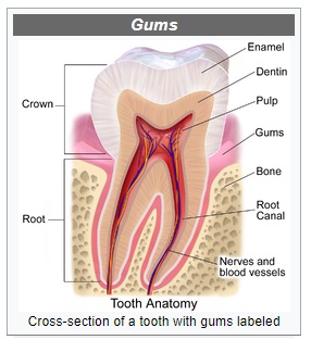
Jump to navigation jump to search. From wikimedia commons, the free media repository. A cross section of a human long bone. Looking at a bone in cross section, there are several distinct layered regions that make up a bone. Bone histomorphometry iwaniec ut, crenshaw td: The outside of a bone is covered in a thin layer of dense irregular connective tissue called the periosteum. Tooth cross section anatomy with all parts including crown neck enamel dentin pulp cavity gums root canal cement bone and blood supply for medical science education and dental health care. The end of a growing tibia, cut lengthwise*. As the names suggest compact bone looks compact and the spongy bone looks like skull bone is a flat bone. A bone is a rigid tissue that constitutes part of the vertebrate skeleton in animals. Virtual bone labwe need our bones to walk, run, jump and move, but this is not all they do. Cross sections and f… leg bones. Advances in fluorescence bone labeling.
Compact bone cross section courtesy: Red marrow fills the spaces in the spongy bone. The outside of a bone is covered in a thin layer of dense irregular connective tissue called the periosteum. Bone marrow is the soft tissue found inside bones that functions mainly to produce red blood cells, white blood cells, and platelets. This simply involves placing a section of the bone on the microscope stage and viewing the.
The wider section at each end of the bone is called the epiphysis (plural = epiphyses), which is filled with spongy bone.
Cross sections and fascial compartmen… category: Draw and label a cross section of a bone. Bone histomorphometry iwaniec ut, crenshaw td: Bones protect the various organs of the body, produce red and white blood cells, store minerals. Cartilages of ear pinnae, epiglottis and eustachian tube. Figure 6.2 long bone short bone irregular bone flat bone sesamoid bone ossification of the epiphysis of a long bone. The wider section at each end of the bone is called the epiphysis (plural = epiphyses), which is filled with spongy bone. The end of a growing tibia, cut lengthwise*. Virtual bone labwe need our bones to walk, run, jump and move, but this is not all they do. A cross section of a human long bone. Cells in different stages of bone growth*. Both types of bone marrow are enriched with blood vessels and capillaries.2. At the outer regions of the section, you can see a dense, thick layer of compact bone.
Cells in different stages of bone growth* cross section of a bone. A bone is a rigid tissue that constitutes part of the vertebrate skeleton in animals.